

Today, in addition to being the Gold Standard diagnostic technique for the investigation of the uterine cavity, hysteroscopy has proven to be an effective and minimally invasive treatment for infertility, early pregnancy loss and chronic abnormal uterine bleeding. Some of the most common uterine pathologic conditions treated by hysteroscopy are polyps, adhesions and fibroids, but there is whole spectrum of uterine pathology that hysteroscopy is the treatment method of choice (see under indications). Even in patients without intrauterine pathology, hysteroscopy can be extremely beneficial as for example, in cases of unexplained repeated implantation failure in IVF.
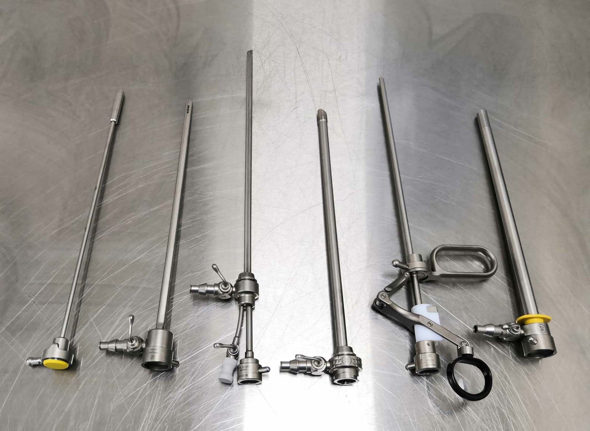
Hysteroscopy allows the direct visualization of the uterine cavity through a rigid, semi‐rigid or flexible endoscope. The hysteroscope consists of a rigid telescope with a proximal eyepiece and a distal objective lens that may be at 0° to allow direct viewing or offset angled at various angles to provide a fore‐oblique view. Advances in fiberoptic technology have led to the miniaturization of the telescopes without compromising the image quality. The total working diameters of modern diagnostic hysteroscopes are typically 2.5 mm to 4.0 mm. Operative hysteroscopy requires adequate visualization through a continuous fluid circulation using an inflow and an outflow channel. The outer diameters of modern operative hysteroscopes have been reduced to a diameter between 4.0 mm and 5.5 mm. The sheath system contains one or two 1.6 mm to 2.0 mm working channels for the insertion of small grasping or biopsy forceps, scissors, myoma fixation instruments, retraction loops, morcellators (surgical instruments used to divide and remove tissue during endoscopic surgery) and aspiration cannula, or unipolar or bipolar electro- diathermy instruments.
Most diagnostic and minor operative procedures can be done in a clinic setting using local anesthesia and fluid distension media, while more complex procedures are generally performed as day surgery under general anesthesia. Operative hysteroscopic procedures require a complex instrumentation setup, special training of the surgeon, and appropriate knowledge and management of complications.

